Karine SERRE, PhD
IMMUNOLOGIST
Latest Posts
21/01/2016
Psoriasis is an autoimmune disease and NOT a chronic inflammatory disease!12/05/2015
CALL For PAPERS: “Innate T Cell Development and Functions in Immune Diseases”28/08/2014
XL Annual Meeting of the Sociedade Portuguesa de Imunologia 13-15th October 201413/04/2013
Beginnings of breaking down the differentiation program of innate IL-17-production27/01/2013
The improbable trio NKG2D – γδ T cells – IgE!!CD8 T cell response to alum-precipitated antigen
CD8 T cells proliferate and produce IFN-g in response to alum-precipitated protein in vivo but do not express Th2-cytokines
Although our knowledge of the diversity in cytokine production by CD8 T cells lags behind CD4 T cells, they too have been reported to fall into two subpopulations based on cytokine secretion so that T cytotoxic type 1 (Tc1) CD8 T cells secrete IFN-g, whereas Tc2 secrete IL-4 (1-5). There is evidence that Tc2 can be induced in vitro by TCR ligation in the presence of IL-4 (3-5), but we questioned whether Tc2 polarization would be achieved in vivo in response to primary immunization with alum-precipitated protein.
This question is of interest because CD8 T cell response to alum-precipitated protein is under debate. Some studies have reported a poor ability of alum-protein vaccines to induce cytotoxic CD8 T cell-responses (6, 7), while we and others have shown that antigen-specific adoptively transferred transgenic CD8 T cells (8) and endogenous CD8 T cells proliferate (9) as well as develop cytotoxicity (10) in response to this form of antigen.
Our approach has been to compare the polarization of transgenic naïve ovalbumin-specific CD4 (OTII) and CD8 (OTI) T cells. This has been done both in their response to TCR ligation in the presence of IL-4 in vitro, as well as during their response to alum-precipitated ovalbumin (alumOVA) in vivo.
Both CD4 and CD8 T cells are polarized to produce Th2 cytokines by IL-4 in vitro
OTI and OTII T cells were activated in vitro using Th2-polarizing conditions. LN cells from OTI or OTII mice were cultured with the OVA-peptide their T cells respectively recognize. Selective Th2-polarization was achieved in both CD8 and CD4 T cells by adding IL-4 with anti-IL-12 and anti-IFN-g.
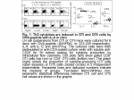
 After 6 days of culture there were many similarities between the responses of the two cell types and the main difference in IL-4 cytokine production was quantitative. Thus medians of 20% of OTII cells and of 2% of OTI cells differentiated into IL-4-producing cells (Fig. 1). Surprisingly IL-5 protein was induced in a median of 1.4% of OTI cells but was never detected in OTII cells. These results clearly confirm that CD8 T cells can be induced to produce IL-4, and thus acquire Th2-features, in response to IL-4-stimulation in vitro.
CD4, but not CD8 T cells, produce Th2 cytokines in vivo in response to alumOVA
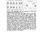
Thus we questioned whether CD8 T cells are also able to acquire Th2 featu res in response to alumOVA in vivo. The responding cells were analyzed for cytokine production after ex vivo restimulation. CD4 T cells were induced to produce Th2 cytokines by this antigen. A subset of 2% of OTII cells differentiating into IL-4-producing effector cells in responses to alumOVA (Fig. 2). This is consistent with other studies assessing the proportion of IL-4-containing OTII cells in response to the same antigen (8, 11, 12). On the other hand, neither IL-4, nor IL-13 and nor IL-5 was induced in vivo in OTI cells responding to alumOVA in LN. Nevertheless, the OTI cells did respond to alumOVA by proliferating and producing IFN-g –a classical feature of Th1 immune responses. These results show that OTI cells responding to alumOVA in vivo fail to mimic the Th2 response of OTII cells responding to this same antigen, but acquire Th1-related features.
Conclusions
The study shows that the Th2-cytokines ‑ IL-4, IL-13, IL-5 (mRNA and/or protein) ‑ as well as the transcription factor GATA-3 (8) are induced in vitro in both OTI and OTII cells when stimulated with OVA-peptide (SIINFEKL or 323-339 respectively) in the presence of IL-4. Although the relative levels of IL-4 expression by responding OTI cells may differ from those induced in OTII cells, in qualitative terms the responses of these two cell types to culture with IL-4 are similar for all other Th2-features. This contrasts with failure of the OTI cells responding to alumOVA in vivo, to mimic the Th2 response of OTII cells responding to this antigen.
These marked differences between the in vivo and in vitro induction of Th2-cytokine production confirm that the in vitro models of Th2 polarization using IL-4 provide at best a poor reflection of Th2 responses in vivo in terms of the diversity of activated T cells produced and the proportion of cells expressing different cytokines. With this in mind we next identified differences in the induction of signaling pathways and transcription regulators in OTI and OTII cells during the in vitro and in vivo responses. The aim of these studies was to obtain clues about molecules involved in CD4 Th2-differentiation signaling during the response to alumOVA in vivo. This has shown that that while subsets of alumOVA-responding CD4 T cells strongly upregulate Th2 and follicular helper T cell features including the surface markers OX40, CXCR5, PD-1, IL-17RB (see more) and the transcription factor c-Maf, CD8 T cells do not (8).
In addition, the proliferative response and IFN-g production induced in CD8 T cells by alum-precipitated protein clearly shows that this form of exogenous antigen is being taken up, processed and effectively presented in association with the MHC class I molecules, a process termed cross-presentation. This is of interest for, despite the poor reported ability of alum-protein vaccines to induce cytotoxic CD8 T cell-responses (6, 7), OTI cells clearly respond to alum-precipitated proteins. This is consistent with previous reports which show that endogenous CD8 T cells proliferate (9) as well as develop cytotoxicity (10) in response to alum-precipitated antigen.
Finally, emergence of “Th2-refractory” IFN-g-producing OTI cells during response to alum-precipitated proteins may have some implication for vaccine designs. Even though the CD8 T cells do not seem to modify the features of the CD4 T cell response studied here, we have found that they have an important impact on the B cell response to the Th2-antigen. During priming with alumOVA, B cells essentially switch to IgG1 (13). However, in the presence of OTI cells the antibody response acquires more Th1-features and B cells switched to a variety of IgG1-, IgG2a- and IgG2b-producers (14). This indicates that the production of IFN-g by CD8 T cells responding to alumOVA has physiological consequences within the overall immune response to alum-precipitated antigen.
References:
1. Cerwenka, A., L. L. Carter, J. B. Reome, S. L. Swain, and R. W. Dutton. 1998. In vivo persistence of CD8 polarized T cell subsets producing type 1 or type 2 cytokines. J Immunol 161:97-105.
2. Carter, L. L., and R. W. Dutton. 1996. Type 1 and type 2: a fundamental dichotomy for all T-cell subsets. Curr Opin Immunol 8:336-342.
3. Croft, M., L. Carter, S. L. Swain, and R. W. Dutton. 1994. Generation of polarized antigen-specific CD8 effector populations: reciprocal action of interleukin (IL)-4 and IL-12 in promoting type 2 versus type 1 cytokine profiles. J Exp Med 180:1715-1728.
4. Sad, S., R. Marcotte, and T. R. Mosmann. 1995. Cytokine-induced differentiation of precursor mouse CD8+ T cells into cytotoxic CD8+ T cells secreting Th1 or Th2 cytokines. Immunity 2:271-279.
5. Noble, A., P. A. Macary, and D. M. Kemeny. 1995. IFN-gamma and IL-4 regulate the growth and differentiation of CD8+ T cells into subpopulations with distinct cytokine profiles. J Immunol 155:2928-2937.
6. Bomford, R. 1980. The comparative selectivity of adjuvants for humoral and cell-mediated immunity. II. Effect on delayed-type hypersensitivity in the mouse and guinea pig, and cell-mediated immunity to tumour antigens in the mouse of Freund’s incomplete and complete adjuvants, alhydrogel, Corynebacterium parvum, Bordetella pertussis, muramyl dipeptide and saponin. Clin Exp Immunol 39:435-441.
7. Garulli, B., M. G. Stillitano, V. Barnaba, and M. R. Castrucci. 2008. Primary CD8+ T-cell response to soluble ovalbumin is improved by chloroquine treatment in vivo. Clin Vaccine Immunol 15:1497-1504.
8. Serre, K., E. Mohr, F. Gaspal, P. J. Lane, R. Bird, A. F. Cunningham, and I. C. MacLennan. 2010. IL-4 directs both CD4 and CD8 T cells to produce Th2 cytokines in vitro, but only CD4 T cells produce these cytokines in response to alum-precipitated protein in vivo. Mol Immunol 47:1914-1922.
9. McKee, A. S., M. W. Munks, M. K. MacLeod, C. J. Fleenor, N. Van Rooijen, J. W. Kappler, and P. Marrack. 2009. Alum induces innate immune responses through macrophage and mast cell sensors, but these sensors are not required for alum to act as an adjuvant for specific immunity. J Immunol 183:4403-4414.
10. Dillon, S. B., S. G. Demuth, M. A. Schneider, C. B. Weston, C. S. Jones, J. F. Young, M. Scott, P. K. Bhatnaghar, S. LoCastro, and N. Hanna. 1992. Induction of protective class I MHC-restricted CTL in mice by a recombinant influenza vaccine in aluminium hydroxide adjuvant. Vaccine 10:309-318.
11. Serre, K., E. Mohr, K. M. Toellner, A. F. Cunningham, R. Bird, M. Khan, and I. C. Maclennan. 2009. Early simultaneous production of intranodal CD4 Th2 effectors and recirculating rapidly responding central-memory-like CD4 T cells. Eur J Immunol 39:1573–1586.
12. Smith, K. M., J. M. Brewer, C. M. Rush, J. Riley, and P. Garside. 2004. In vivo generated Th1 cells can migrate to B cell follicles to support B cell responses. J Immunol 173:1640-1646.
13. Mohr, E., K. Serre, R. A. Manz, A. F. Cunningham, M. Khan, D. L. Hardie, R. Bird, and I. C. MacLennan. 2009. Dendritic cells and monocyte/macrophages that create the IL-6/APRIL-rich lymph node microenvironments where plasmablasts mature. J Immunol 182:2113-2123.
14. Mohr,E., A.F. Cunningham, K.M. Toellner, S. Bobat, R. Coughlan, R. Bird, I.C.M. MacLennan and K. Serre. 2010. IFN-g produced by CD8 T cells induces T-bet-dependent and independent class switching in B cells in responses to alum-protein vaccine. Submitted