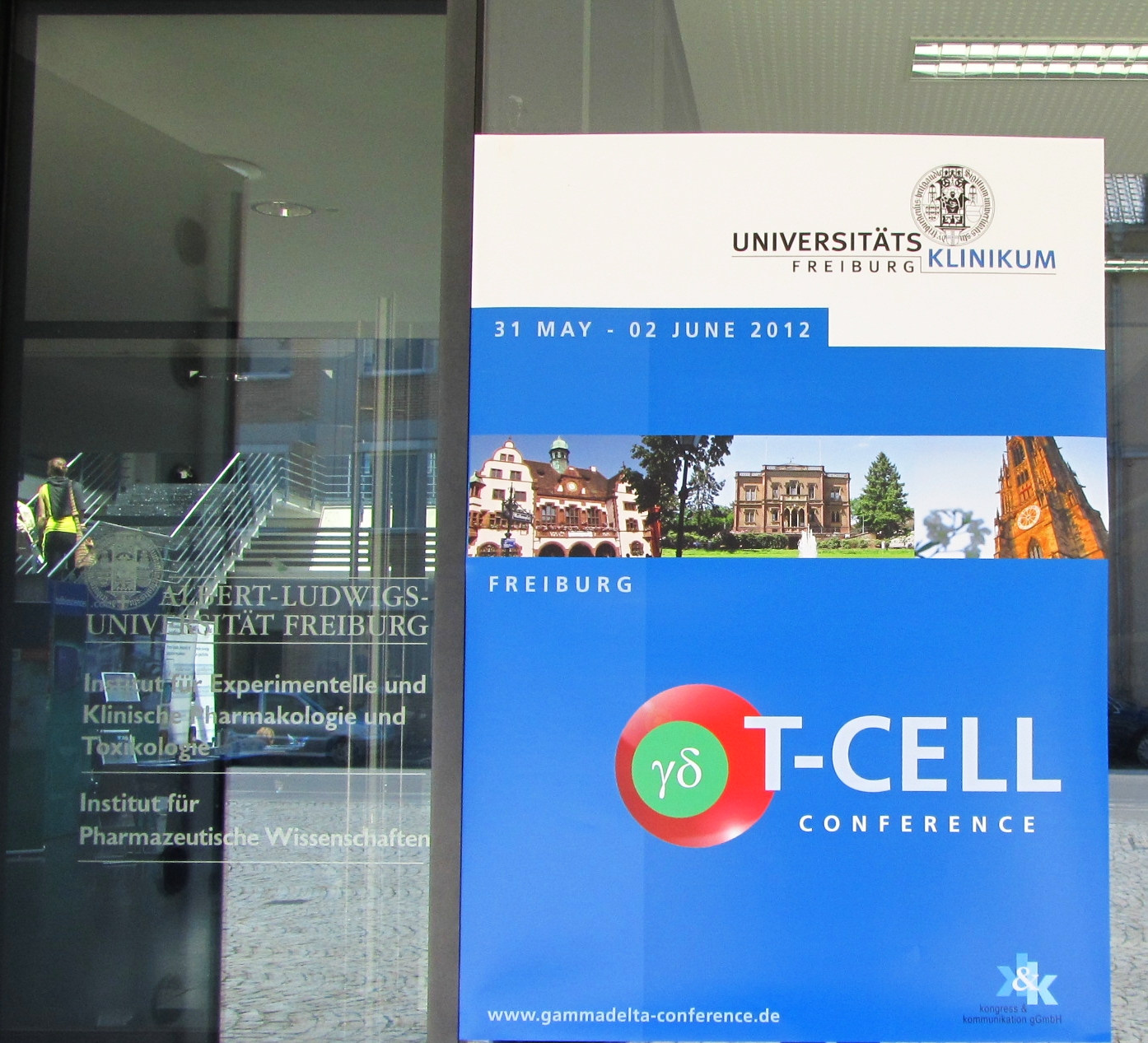
γδ T cell conference report Freiburg 2012
Back from the conference, filled up with news about the novel hot topics in the field and with challenging discussions with some of the currently leading researchers on the ever growing surprising γδ T cells!
But before, just to let you know, Freiburg (im Breisgau), nicely situated in the south of Germany some 50 km away from Basel, is a very friendly city, with a cute centre and a charming street market
next to the main cathedral!
We were lucky to enjoy overall very good weather and a walk up to a pretty view-point over the city.
Now some science, so here is a short report on what was said (more especially on murine γδ T cells, I have to admit).
• Sessions A-B-C –> γδTCR antigens, mechanisms of recognition, T cell activation
Thus, during the first day we have been reminded how little we understand about what are the γδTCR ligands. However, some very interesting talks have shed new lights on the subject and offered some novel interesting potential candidates. Thus, now new γδTCR antigens can be added to the non exhaustive list of non-classical major histocompatibility complex class I molecules T10 and T22, the MHC class I-like molecules MICA and MICB, the ATP synthase-F1 (AS)/apolipoprotein A-1 complex, non-peptidic pyrophosphomonoesters (phospho-antigen), etc…
– Ben Willcox (University of Birmingham, UK) convincingly showed that the Endothelial Protein C Receptor (EPCR) is directly recognized by a Vγ4Vδ5 human clone. Although EPCR has a MHC-like structure related to CD1d and that it binds lipids, they showed that EPCR-TCR interaction is lipid-independent. Interestingly, EPCR is up-regulated on cytomegalovirus (CMV) infected cells, and on tumour cells.
– In a very fruitful collaboration with Julie Dechanet-Merville (Bordeaux Segalen Univesité, France), they also characterized a ligand of non-Vδ2 γδ T cells that specifically expand during CMV infection. These non-Vδ2 γδ T cells generated during infection are usually able to kill tumour cell lines suggesting they recognise self-antigens. Using various approaches they show that the Erythropoeitin-Producing Hepatocyte kinase A2 (Ephrin receptor A2 = EphA2), a member of the largest family of tyrosine kinases receptor is recognised by Vγ9Vδ1 cells. Interestingly, they even suggest a shared reactivity towards EphA2 may exist within the Vδ1 T cells. Again, EphA2 is up-regulated on CMV-infected cells, and during tumogenesis.
– In line with Ben Willcox’s talk Erin Adams (Chicago, USA) provides structural evidence that there is a small but significant population of human γδ T cells that respond to MHC-like protein CD1d when presenting the glycosphingolipid sulfatide. Theses sulfatides represent about 20% of myelin glycolipids, thus, it would be interesting to assess the response and proportion of this γδ T cell population during multiple sclerosis.
– No more MHC-like molecules :] with Anne Kaiser (Freiburg, Germany) who has data that a Vγ9Vδ2 TCR clone recognize the human HLA B58 alloantigen. This γδ T cell clone does not recognize Daudi or phosphoantigens, and does not express NK receptors.
– Jessica Bruder (Munich, Germany) has elegantly identified aminoacyl-tRNA-synthetase as the ligand of a pathogenic Vγ1.3Vδ2 clone isolated from a patient with polymyositis.
– Willi Born (Denver, USA) has provocative results indicating that just as like diabetogenic ab T cells in NOD mice, γδ T cells directly bind to an insulin peptide 9-23. However, γδ T cells cannot completely follow ab T cell rules and, there is a difference as they do not require APC! Instead they directly bind to this peptide when it is oxidixed in its cysteine (position 19) and thus forms dimers.
– Two very interesting talks from the same lab Emmanuel Scotet and Steven Nedellec (Nantes, France) provide evidence that CD277, which is a member of the B7 receptor family related to butyrophilins, plays a role in human γδ T cell activation. Indeed, agonist antibody specific for CD277 specifically activates human peripheral Vγ9Vδ2 T cells and induces cytolysis, CD69 expression, IFN-γ and TNF-α production, and proliferation in a TCR-dependent manner. Even if they show it is cell-to-cell dependent, the mechanism of action of CD277 still remains to be elucidated as it is not known if there is a need for phospho Ag in this interaction or if the TCR directly interacts with CD277.
– Again two related talks from Thomas Zal and Grzegorz Chodaczek (Houston, USA) who presented their very nice data from Nature Immunology 13, 272–282 (2012) paper. Body-barrier surveillance by epidermal γδ TCR. This suggests for the first time that homeostatic maintenance of mouse dendritic epidermal T cells (DETCs) requires constant γδ TCR engagement, as visualized by the strong staining obtained with confocal microscopy on skin tissue section with anti-CD3zeta phosphorylated tyrosine 142. The epidermal ligand recognized by the DETC is still unknown. Thus, intraepidermal T cells are truly autoreactive, constitutively transmitting signals from a putative ligand expressed by normal epithelial cells, from conventional or germ free mice.
– Staying with DETC, a provocative talk from Gleb Turchinovich (London, UK) who shows that murine Vγ5Vδ1 T cells that are selected on a mutated Skint-1 molecule (from mice from Taconic) are “tolerised”. Indeed, Vγ5Vδ1 T cells have defect in proliferation, phosphorylation of ERK and calcium signalling when re-stimulated in vitro with anti-CD3. Importantly the state of anergy is imprinted during thymic development. I would have liked to see the precise γδTCR affinity between “Taconic” and “Jackson” Skint-1 molecules.
– Finally, E Dopfer (Freiburg, Germany) and Balbino Alarcon (Madrid, Spain) gave us information on the importance of conformational changes on the CD3 molecules that plays crucial role in both γδ and ab T cell activation.
In conclusion, overall γδ T cells can all be seen as auto-reactive cells sensitive to “stress-reporter” self-molecules. Most of the time these cells do not require any quid of processing, but are recognised directly by the γδTCR. On one hand, γδ T cell activation can occur through direct up-regulation of one or many of these newly characterized stress-self-molecules, and on the other it can happen in the context of a stress-related increase of costimulatory molecules in the micro-environment.
• Sessions D-E-F –> γδ T cell development, phenotype, and function at steady state
–> Suffice to say that, in this year conference, most of the attention is focussed on IL-17-producing γδ T cells (γδ17) and less on the IFN-γ-producing γδ T cells (γδ1)!
One of the first questions was: Is the TCR-signal strength an important regulator of development of γδ17? Although the development of γδ T cells relies on strong TCR signal during selection, it was proposed that strong TCR signalling favours commitment to γδ1 over γδ17. Two independent labs, using completely different systems, provided contradictory results on this subject.
– Nital Sumaria (London, UK) identified a population of IL-17+CD27+ γδ T cells from 3 days post-natal thymus. This is the first time that IL-17 secretion is detected in CD27+ γδ T cells. This also shows that commitment to IL-17A is acquired early in the thymus, and this is consistent with the embryonic wave of γδ17 cell production (see Jan Haas).
Interestingly, during in vitro culture in the presence of OP9-DL1 for 7 days, IL-17+CD27+ γδ T cells would give rise to 80% CD27- / 20% CD27+ cells, while IL-17-CD27+ γδ T cells would give rise to 50% CD27- / 50% CD27+ cells. This raises the question of the progenitors for each CD27+ and CD27- γδ T cell subsets.
Then, CD25+CD27+ γδ T cells were cultured with GL3 Ab giving rise to about 100% CD25-CD27+ γδ T cells, suggesting TCR-engagement especially favours the differentiation towards CD27+ T cells.
To further assess this, they use a different model where γδ T cells lack both extracellular variable Ig-like domains and thus cannot engage any ligand. Surprisingly, in vitro results show no advantage for CD27- γδ T cells in this system, and the differentiation of about 85% of γδ T cells that express CD27!
This demonstrates for the first time that CD27+ γδ1 T cells can be generated in the complete absence of TCR-signalling.
– By contrast, Melanie Wencker (London, UK) uses a completely different in vivo approach, by examining the phenotype of SKG mice (BALBc) that have a mutation in ZAP70 leading to attenuated TCR-signaling. Counter intuitively, these mice have less CD27-CD44++ γδ17 T cells both in adult thymus and secondary lymphoid organs. The proportion L-17-competent cells is already greatly reduced in embryonic thymic lobes. In addition, in vitro restimulation (I cannot remember the stimulus…) led to a decrease in the induction of IL-17-producing γδ T cells.
These mice suffer from arthritis but in a manner that is dependent on CD4 Th17 cells!! Therefore, it would be important to see if naïve CD4 T cells, from SKG mice, respond normally to Th17-conditioning culture polarization. The result may stress again the difference between ab T cells and γδ T cells.
As these results are contradictory to previously published data suggesting that γδ17 cells are less susceptible to TCR-signalling, and given the differences observed between Taconic and Jackson mice, we may need to re-evaluate the balance in γδ1 / γδ17 developmental in pre-commitment in different strains of mice.
–> Two talks highlighted the importance of two cytokines (IL-7 and IL-17 itself) in controlling development and maintenance of IL-17-producing γδ T cells in vivo.
– Marie Laure Michel (London, UK) proposes to add IL-7 to TGF-β and IL-6 cytokines that control the thymic development of γδ17 cells. The γδ17 cells are characterized by CD44+IL-7Rα and CD27-. In vitro cultures of γδ T cells in the presence of IL-7 induce the selective expansion of IL-7Rα+ CD44+ RORyt+IL-17+ cells.
To assess potential role of IL-7 in vivo mice were induced psoriasis that increases the proportion of IL-17-secreting γδ17 cells. Treatment with anti-IL-7R reduced the development of IL-17-secreting γδ17 cells from 16% to 6% and favoured the generation of IFN-γ-secreting γδ1 cells from 40% to 54%. IL-7 acts through STAT3 and control the proliferation of the γδ17 cells. This property is also true for human γδ cells (PBMC or cord blood), which respond to IL-7 in the presence of anti-TCR, with a 20-folds increase in IL-17-secreting γδ17 cells.
– Jan Haas (Freiburg, Germany) presented his “soon-to-be-published” elegant story on the development of γδ17 cells that occurs essentially during a small window between day E16.5 to E18.5, and then stopped after birth. Therefore, γδ17 cells are predetermined in the embryonic thymus and persist as long-lived and self-renewing cells in adult mice. IL-17 itself plays a negative feedback and blocks the development of γδ17 cells perinatally. However, the active source of IL-17 in this system needs to be identified as they include innate lymphoid cells, CD4 Th17 cells, NK cells, NK T cells.
– Julie Jameson (La Jolla, USA) showed that obesity induces a drastic decrease in Vγ5 DETC, while the proportion of Langerhans cells remains constant, IEL and γδ T cells in intestine.
–> Finally, I was really enthusiastic by two special talks.
– Leo Lefrançois (Connecticut, USA) provides exciting results about immune response induced by immunization with Listeria monocytogenes. They generated genetically modified Lm that expresses the ligand for mouse E-cadherin. After oral challenge the proportion of CD27- CD44++ Vγ6Vδ1 γδ17 T cells in mesenteric LN, goes from day 0 naïve 0.6% to 50% day 10, and remain constant at 40% for up to day 120. Interestingly, there is increase in the CD27- CD44++ γδ17 T cell subset, with each challenge suggesting a “memory” phenotype. To my surprise, the CD27- CD44++ T cells contain effectors that secrete IL-17A, IFN-γ and both cytokines.
I would have liked to know if IL-1β, IL-23, IL-6 or TGF-β are required for the generation or/and emergence of this CD27- CD44++ T cell population. Nevertheless, a very challenging talk indeed!
– Thomas Boehm (Connecticut, USA) asks a very complex question that is: “what is the minimal number of factors required to make a functional thymus?”
And to address this, the approach is to try to rebuild a thymus in a nude mouse. Nude mice are deficient in Foxn1. Thus, they analysed the development of T cells in thymic-enforced expression of various genes. This shows that:
Scf + CCL25 –> give rise to Mast cells.
Scf + CCL25 + CXCL12 –> give rise to B cells
Scf + CCL25 + DLL4 –> give rise to DN cells
Scf + CCL25 + CXCL12 + DLL4 –> give rise to DP cells
Cool stuff no? Soon-to-be-published I think!
• Sessions G-H –> γδ T cells in infectious and autoimmunity diseases
– RL O’Brien (Collorado, USA) has interesting data about C57BL10 female mice that develop spontaneous keratitis (corneal inflammation).
Rederived mice develop similar disease than normal mice, suggesting that there is no involvement of infectious agent that could be congenically acquired in the strain.
Depletion / inactivation of B cells with long-term administration of an anti-CD79 do not change the indidence of the disease, suggesting that neither antibodies nor B cells contribute to the disease.
Transfer of T cells from mice suffering form keratitis, transmit the disease, suggesting an involvement of T cells.
Transfer of Vγ1 γδ T cells, into TCRd-/- reduced severity and incidence of disease. Of note Vγ4 T cells do not protect from disease.
Female hormones do not explain why only females get the disease.
B10 mice have more tear in their eyes and yet have more disease incidence demonstrating that dry eye do not increase the disease.
– Vica Marcu-Malina (Tel Hashomer, Israel) studied juvenile (less than 16 years old) idiopathic (cause unknown) arthritis (inflammation of the joint), and assessed the role of γδ T cells.
In response to IPP Vγ9 T cells produce TNF-α and IFN-γ.
Vγ9 T cells from synovia are more activated than Vγ9 T cells from blood.
The production of IPP occurs in joint via synovial fibroblasts. Indeed, Vγ9 T cells cultured with synovial fibroblasts up-regulate CD69 and produce IFN-γ.
However, Vγ9 T cells from synovia proliferate less than Vγ9 T cells from blood in response to IPP. This is in part due to the presence of more Foxp3+CD4+CD25+ in the synovium.
Interestingly, Vγ9 T cells activated via IPP kill / destruct synovial fibroblasts.
Taken all together these data demonstrate a pathogenic role of γδ T cells during JIA.
– S Pantelyushin (Zurich, Switzerland) has interesting data on psoriasis in mice. This was recently published in J Clin Invest. 2012 May1. Rorγt+ innate lymphocytes and γδ T cells initiate psoriasis form plaque formation in mice.
Mice were shave and creamed with Aldara (Imiquimod = TLR7/8 agonists). Of note, this cream is usually used on skin to treat superficial basal cell carcinoma in adults with normal immune systems. Six day after treatment mice develop skin psoriasis.
Treatment with anti-IL-23p40 is protector as well as anti-TNF-α, although to a lesser extent. The cytokine signature includes IL-17a and IL-22 production by γδ T cells. RORyt-GFP shows that the principal cytokine producers are γδ T cells. Vγ4 T cells are the main cause of the inflammation. In the absence of γδ T cells ILC cause the Aldara-induced psoriasis disease.
• Sessions I-J –> γδ T cells in cancer: Foes or Friends?
– Telma Lança (Lisboa, Portugal) proposes an essential role for the CCR2-CCL2 chemokine axis in the migration of γδ T cells to the tumour. Importantly, in the mouse model of B16 melanoma γδ T cells are shown to be protective, while they are pro-tumoral in other kind of cancers.
This is autopromo :] since Telma is from Bruno Silva-Santos’ lab.
General conclusions:
The next venue in 2014 should be Chicago thanks to Professor ZW Chen.
Overall I really enjoyed the meeting 8D.
My only advice for the 6th gamma-delta T cell conference would be to take more attention to the poster sessions and find a way to have all the posters presented at all times during the conference. Having poster sessions with wine and cheese/nibbles would be in fact perfect to help people discussing in front of the posters, but may be this is too much to ask ;]
In any case, hopefully see you all in two years time!!


